
This course meets the need for obstetricians or radiologists with advanced training in diagnostic imaging. The course combines theoretical-practical online modules and on-site training in Barcelona. The first part includes online training with modules covering basic and advanced knowledge acquisition, along with various practical tasks and image submissions to validate the accreditation of these competencies.
Language: English ![]()
Directors
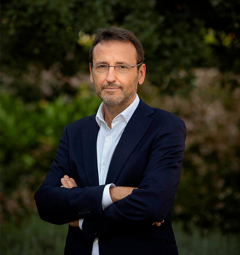 Eduard Gratacós
Eduard Gratacós is the director of BCNatal (clinical center) and BCNatal-FMRC (research).
Expert and pioneer in fetal medicine and surgery, he has participated in the design and first application of several fetal surgery procedures. He has made significant scientific contributions in different fields, including fetal growth restriction and preeclamsia, fetal surgery and monochorionic twins.
He is the author of more than 600 peer-reviewed articles and several patents. Has trained over 400 doctors in fetal medicine and surgery, and directed over 40 doctoral theses.
He has coordinated 60+ and participated in 75+ international and national research projects funded by national and international research agencies.
Former Editor-in-Chief of fetal Diagnosis and Therapy and Scientific Chair of ISUOG.
Founder of Fetal I+D Barcelona, which offers international medical training in collaboration with various universities, with over 25,000 users worldwide.
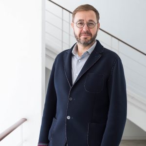 Francesc Figueras
Head of the Department of Fetal-Maternal Medicine at Hospital Clinic (Barcelona) and Full Professor of the University of Barcelona.
Master Degree in Health Methodology by the University Autonomous of Barcelona.
Postgraduate degree in Public Healt by the University of Edinburg.
Research interest in Fetal Growth Restriction and Preeclampsia: Author of ~260 articles in indexed journals (~760 Impact Factor). H-Factor 59. Author of 15 chapters in books & Editor of 6 books.
Invited speaker in more than 200 international congresses and conferences.
Director of 14 PhD Thesis Projects.
Principal Investigator of 10 public-funded competitive research projects.
Professors
 Mar Bennasar
Mar Bennasar is a Materno-fetal consultant of the Materno-Fetal department at BCNatal (Hospital Clínic and Hospital Sant Joan de Déu of Barcelona).
She is specialized in fetal cardiology and fetal therapy with more than 15 years of experience.
She has published more than 40 scientific papers in international journals. She usually participates in national and international courses and congresses as a faculty member.
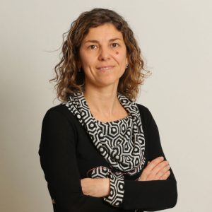
 Olga Gómez
Since 2008, Olga Gómez is a member of the Fetal Pathology and Cardiology Units at BCNatal, a referral center in Maternal-FetalMedicine at the Hospitals Clinic and Sant Joan de Déu in Barcelona, and of the Fetal Medicine Research Center (Fetal i+D), a multidisciplinary team with one of the largest scientific international productions.
She is currently the coordinator of the Fetal Cardiology unit, one of the largest reference units for the evaluation of cardiac malformations in Spain, with more than 100 major confirmed cardiac defects yearly. With a broad background in fetal medicine and particularly in advance fetal ultrasound and echocardiography, in recent years her research has focused on the functional echocardiographic evaluation of congenital heart disease. In 2014, she spent a year as a research fellow in the Pediatric Cardiologist Division at Sickkids Hospital in Toronto Canada.
Olga Gómez is also an associate Professor of Obstetrics at the University of Barcelona. She has participated as a speaker in more than 70 courses and seminars on fetal pathology and echocardiography and has been involved in different clinical research projects. As a result of her professional activity she has authored more than 65 papers published in indexed journals and she has directed two doctoral theses.
 Miriam Illa
Medical specialist in Fetal Medicine at BCNatal (Hospital Clínic de Barcelona and Hospital Sant Joan de Déu).
Post-doctoral researcher and teacher in numerous specialized courses in Fetal I + D Education and international conferences.
She is the moderator of the forum “Normal pregnancy”.
 Marta López
Consultant doctor of the Maternal and Fetal Medicine Department in Hospital Clínic of Barcelona. Doctor in Medicine from the University of Barcelona in 2015.
Associate Professor in the degree of Medicine of the University of Barcelona.
IDIBAPS researcher in the “Research in Fetal Medicine and Perinatology” team.
Member of the Perinatal Infections Unit and the Maternal High-Risk Obstetrics Unit. Specialist in the management of maternal or TORCH infections during pregnancy and in the control of pregnant women with maternal heart disease, oncological pathology and placental pathology.
She has published scientific articles in international journals with impact factor. She has participated as a speaker in national and international courses and congresses in the field of perinatal infections and maternal pathology.
 Narcís Masoller
Narcís Masoller is a senior specialist in the Maternal-Fetal Medicine service at Hospital Clínic de Barcelona.
He focuses most of his daily activity in the Units of Fetal Cardiology, Fetal Neurosonography and in the Unit of Placental and Myometrial Pathology.
Doctor of Medicine from the University of Barcelona, he has published more than 20 scientific articles in international journals with an impact factor and he has participated as a teacher in numerous courses and conferences.
 Edurne Mazarico
She is a Doctor in Medicine and Surgery and specialist in Gynecology and Obstetrics.
She works at Hospital Sant Joan de Déu, University of Barcelona. She combines clinical care with teaching and research.
She is responsible for the Intrauterine Growth Restriction and Preeclampsia Unit of the Hospital Sant Joan de Déu.
In addition, she is a researcher at the Maternal and Child Health and Development Network (RedSAMID) funded by the Carlos III Institute of Health.
She collaborates in the research lines “Fetal environment and obstetric complications” and ” Growth Restricion and Preeclampsia” of BCNatal.. He carries out his research in collaboration with the San Joan de Déu Foundation and the Fetal Medicine Barcelona Foundation (Fetal I D).
 Anna Peguero
Senior Specialist in Maternal-fetal Medicine of the Hospital Clínic de Barcelona.
Co-responsible for the Obstetric Intensive Care Unit and the Delivery Room. Member ot the Intrauterine Growth Restriction and Preeclampsia Unit. Head of the Perinatal Mortality Committee.
Specialized in the management of severe obstetric pathology and in the control of pregnant women with hypertension and poor obstetric history.
She has published scientific papers in international journals with impact factor, especially in the field of preeclampsia and placental pathology.
 Miriam Pérez
Postdoctoral researcher and teacher in numerous specialized courses in Fetal I + D Education and international conferences.
She is a researcher for the Maternal and Child Health and Development Network (RedSAMID) funded by the Carlos III Health Institute and focuses her research on neurodevelopment in congenital heart disease.
She is responsible for the prenatal circuit of Perinatal Palliative Cures group at the Sant Joan de Déu Hospital.
 Joan Sabrià
Consultant in the Fetal Medicine Unit at BCNatal (Hospital Sant Joan de Déu y Hospital Clínic de Barcelona) and Associated Professor at the University of Barcelona Medical School.
Specialized in Prenatal Diagnosis and Fetal Pathology with more than 15 years of experience.
Has authored more than 30 peer reviewed international scientific publications and usually participates in national and international courses and congresses as a faculty member. Past member of the Catalan Chapter of Fetal Medicine and member of the Catalan Ultrasound Screening Committee for the Catalan Health Department.
 Iris Soveral
Degree in Medicine from the Autonomous University of Barcelona (2014-2010).
Specialty in Obstetrics and Gynecology at the Hospital Clínico de Barcelona (2011-2015).
Awarded the Josep Font Residency Award granted by the Hospital Clínico de Barcelona for the development of a research project in the field of Congenital Heart Disease in Prenatal Life.
In this context, I was clinical fellow Echocardiography and Fetal Pathology at BCNatal, with clinical stays also in the Adult Congenital Heart Disease unit of the Hospital Clínico de Barcelona and in Pediatric Cardiology at the Hospital Sant Joan de Déu in Barcelona.
Doctorate from the University of Barcelona (Excellent Cum Laude) in 2021.
Presently, I am especially interested the fields of Fetal Medicine, Reproductive Genetics and Prenatal Diagnosis, particularly early prenatal screening and prenatal application of chromosomal microarray analysis and exome sequencing. Currently working as a specialist in Maternal and Fetal Pathology.